OVERVIEW
NEUROANATOMY
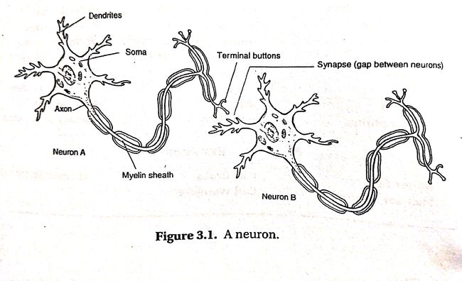
Neuroanatomy refers to the study of the parts and function of neurons
Dendrites(树突) - Rootlike parts of the cell that stretcdh out from the cell body
Cell Body(Soma) - Contains the nucleus and other parts of the cell needed to sustain its life
Axon - Wirelike structure ending in the terminal buttons that extends from the cell body
Myelin Sheath - A fatty covering around the axon of some neurons that speeds neural impulses
Terminal Buttons(End Buttons, Terminal Branches of Axon, and Synaptic knobs) - The branched end of the axon that contains neurotransmitters
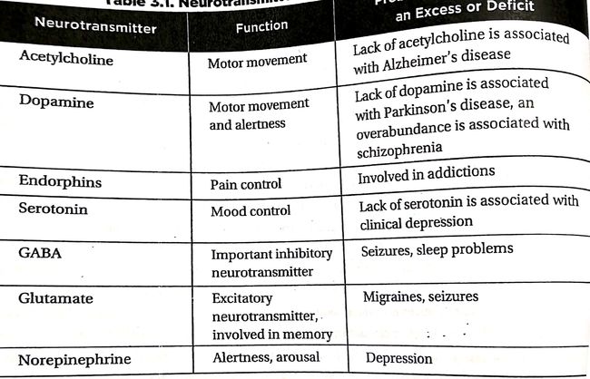
Neurotransmitters(神经递质) - Chemicals contained in terminal buttons that neurons to communicate
Synapse(突触) - The space between the terminal buttons of one neuron and the dendrites of the next neuron
How a Neuron "Fires"
和生物课上讲的一样
All-or-none Principle - A neuron either fires completely or it does not fire. It depends on whether the Neurotransmitters are accumulated to threshold.
Neurotransmitters
Some neurotransmitters are Excitatory, meaning that they inhibit the next cell from firing. Other neurotransmitters are Inhibitory, meaning that they inhibit the next cell from firing
NERVOUS SYSTEM
Afferent Neurons (or Sensory Neurons) - Neurons that take information from the senses to the brain
Interneurons - Neurons that pass information from Afferent Neurons to elsewhere in the brain or on to Efferent Neurons
Efferent Neurons (or Motor Neurons) - Neurons that take information from the brain to the rest of the body
Organization of The Nervous System
The Central Nervous System
Our brain and spinal cord
The Peripheral Nervous System
All other nerves in our body - all the nerves not encased in bone. Two categories: The Somatic and The Autonomic Nervous Systems
Somatic Nervous System - controls our voluntary muscle movements. Controlled by moter cortex of the brain
Autonomic Nervous System - controls the automatic functions of our body-our heart, lungs, internal organs, glands, and so on. control our response to stress. Two categories: The Sympathetic and Parasympathetic Nervous Systems
Sympathetic Nervous System - mobilizes our body to respond to stress. Carry messages to organs, glands, and muscles that direct our body's response to stress. The alert system of our body. Accelerates some functions (such as heart rate, blood pressure, and respiration) but conserves resources needed for a quick response by slowing down other functions(such as digestion)
Parasympathetic Nervous System - Slow down our body after a stress response
Normal Peripheral Nervous System Transmission
Example:
- Stub your toe on a cast-iron coffee table
- Sensory neurons in your toe are activated
- Message transmitted to spine(Afferent Nerves)
- Message goes through spine into your brain and your Sensory Cortex
- Motor cortex sends information down the spine cord to muscles control legs and foot(Efferent Nerves)
- You hold your foot
Reflexes: An Important Exception
Reflexes do not obey the above process.
Eg. Knee-jerk Reaction. Response to intense heat or cold
THE BRAIN
Ways of Studying the Brain
Accidents - Part of the brain of a patient is damaged and the doctor is able to study the patients behavior after the damage
Eg. In 1848, a railroad worker named Phineas Gage was involved in an accident that damaged the front part of his brain. Gage's doctor took notes documenting the brain damage and how Gage's behavior and personality changed after the accident. Gage became highly emotional and impulsive after the accident. Researchers concluded that the parts of the brain damaged in the accident are somehow involved in emotional control
Lesions - Lesioning is the removal or destruction of part of the brain. It is usually not done for research purpose. Some patients have no choice but to remove part of the brain and doctors will monitor the patient after the surgury.
Eg. In the past doctors control mentally ill patients by frontal lobotomy. Lesioning part of the frontal lobe would make the patients calm and relieve some symptoms. Drugs replaced frontal lobotomies nowadays
Electroencephalogram(EEG) - Detect brain waves. Used to examine different stages of conciousness. It is widely used in sleep research to identify the different stages of sleep and dreaming
Computerized Axial Tomography(CAT or CT) - Use several X-ray cameras rotate around the brain and combine all the pictures into a detailed three-dimensional picture of the brain's structure. It is used to look for brain tumor
Magnetic Resonance Imaging(MRI) - MRI and CT both give pictures of the brain. MRI use magnetic field to measure the density and location of brain material. Bot MRI and CT only gives the structure of the brain
Positron Emission Tomography(PET) - Measure which area of the brain is most active when certain tasks are performed. It measures how much of a certain chemical parts of the brain are using. Different types of scan are used for different chemicals
Functional MRI - New technology which combines MRI and PET. Show details of brain structure with information about blood flow in the brain.
Brain Structure and Function
Three separate major categories or sections: the hindbrain, midbrain, and forebrain
HINDBRAIN
Controls basic biological functions
Medulla(medulla oblongata) - control our blood pressure, heart rate, and breathing. Located above the spinal cord
Pons - Connects the hindbrain with the midbrain and forebrain. Involved in the control of facial expressions. Located above the medulla and toward the front
Cerebellum - Coordinates some habitual muscle movements, such as tracking a target with our eyes or playing the saxophone. Located on the bottom rear of the brain
MIDBRAIN
This area is between the hindbrain and the forebrain and integrates some types of sensory informationand muscle movements
Reticular Formation - A netlike collection of cells throughout the midbrain that controls general body arousal and the ability to focus our attention. We will fall into deep coma if this area does not function
FOREBRAIN
Control what we think of as thought and reason. Most psychological researchers concentrate on this area
Thalamus - Receive sensory signals from spinal cord and send to other parts in the forebrain. Located on top of the brain stem
Hypothalamus - Controls several metabolic functions, including body temperature, sexual arousal (libido), hunger, thirst, and the endocrine system. Located right next to the thalamus
Amygdala and Hippocampus - Amygdala is vital to our experiences of emotion, and the hippocampus is vital to our memory system. Memories must pass through this area in order to be encoded, otherwise people cannot retain new information
Cerebral Cortex
The layer covering the rest of our brain which has fissures
Hemispheres
Contralateral Control - Two hemispheres control the other side of the body. Left controls right, right controls left
Brain Lateralization or Hemispheric Specialization - Possibility that left hemisphere may be more active during logic and sequential tasks and the right during spatial and creative tasks. This generalization requires further researches before conclusions are drawn
Split-brain Patients - Patients whose Corpus Callosum is cut in order to treat severe epilepsy.
- Researchers study Brain Lateralization by studying Split-brain Patients
- Split-brain Patients cannot orally report information on right hemisphere since the spoken language centers of the brain are usually located in the left hemisphere
Areas of the Cerebral Cortex
Association Area - Any area of the cerebral cortex that it is not associated with receiving sensory information or controlling muscle movements
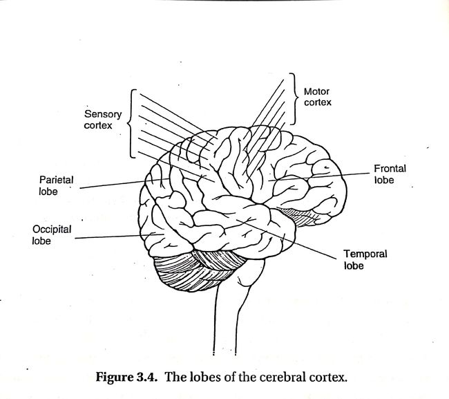
FRONTAL LOBES
Large areas od the cerebral cortex located at the top front part of the brain behind the eyes
Prefrontal Cortex - Located at the front of the front lobe. Plays a critical role in directing thought processes. Is responsible for abstract thought and emotional control
Broca's area - Usually located at the left hemisphere of the frontal lobe. Responsible for controlling the muscles involved in producing speech. Damage of this area may cause us unable to move the muscle needed for speech
Motor Cortex - Located at the back of frontal lobe. Control voluntary movements. The bottom of this area controls the top part of the body and vice versa
PARIETAL LOBES
Located behind the frontal lobe but still on the top of the brain
Sensory Cortex(somato-sensory cortex) - Located right behind the motor cortex. Receive incoming touch sensations from the rest of our body. The bottom of this area receive information from the top of the body and vice versa
OCCIPITAL LOBES
Located at the back of the brain. Interpret messages from the eyes
TEMPORAL LOBES
Process sounds from our ears. It is not lateralized
Wernicke's Area - Interpret both written and spoken speech. Damage to this area would affect our ability to understand language. Speech might sound fluent but lack proper syntax and grammatical structure
Brain Plasticity
- If part of the brain is damaged, the other part would make new connections and take over the function of the damaged part
- Younger brains are more plastic
ENDOCRINE SYSTEM
Adrenal Glands - Produce adrenaline which signals the rest of the body to prepare for fight or flight
Ovaries and Testes - Produce sex hormones
Genetics
Basic Genetic Concepts - pass
Twins - pass
Chromosomal Abnormalities - pass


