利用深度学习对医学CT图像(LIDC-IDRI)中的肺结节进行良恶性判断
肺癌是最常见的癌症,目前,CT可用于帮助医生在早期阶段检测肺癌。 在许多情况下,识别肺癌的诊断取决于医生的经验,这可能会忽略一些患者并导致一些问题。 在许多医学影像诊断领域,深度学习已被证明是一种流行且有效的方法。 本文主要基于LIDC-IDRI这一公开数据集,对其进行了肺结节的提取,并利用CNN对其分类训练,从而辅助医生作出判断。
由于篇幅较长,将分为2篇博客,这篇主要介绍数据处理,即肺结节的提取。
数据集
数据集采用为 LIDC-IDRI (The Lung Image Database Consortium),该数据集由胸部医学图像文件(.dcm)(如CT、X光片)和对应的诊断结果病变 标注(.xml) 组成。数据是由美国国家癌症研究所(National Cancer Institute)发起收集的,目的是为了研究高危人群早期癌症检测。
该数据集中,共收录了1018个研究实例。对于每个实例中的图像,都由4位经验丰富的胸部放射科医师进行两阶段的诊断标注。在第一阶段,每位医师分别独立诊断并标注病患位置,其中会标注三中类别:
- 大于等于3mm的结节
- 小于3mm的结节
- 大于等于3mm的非结节
在随后的第二阶段中,各位医师都分别独立的复审其他三位医师的标注,并给出自己最终的诊断结果。这样的两阶段标注可以在避免forced consensus的前提下,尽可能完整的标注所有结果。
图像信息(.dcm)
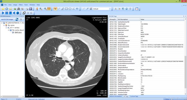
图像文件为Dicom格式,是医疗图像的标准格式,其中除了图像像素外,还有一些辅助的元数据如图像类型、图像时间等信息。
一张CT图像有 512x512 个像素点,在dicom文件中每个像素由2字节表示,所以每张图片约512KB大小。
目前测试一共1012个病例数据,对于每个实例,可以看为一个三维矩阵D(slicer * rows * cols), slicer表示切片的个数(对应每个病例的.dcm文件数),rows和cols分别表示图片的行数和列数(默认为512)。
eg: 对于病例LIDC-IDRI-0001,即为 133 × 512 × 512 133 \times 512 \times 512 133×512×512的矩阵,一共133张切片,每张大小 512 × 512 512 \times 512 512×512。
查看dcm文件:
- 通过pip或者Anaconda安装pydicom模块,该模块是python专门用来处理dicom格式文件的库。
通过上面2种方式,我们可以看出dicom文件中包含了一些图像信息(SOP Instance UID、Study Instance UID,Series Instance UID······)
SOP Instance UID: 用于唯一区分每一张dcm切片
Study Instance UID: 每个病例对应的检查实例号
Series Instance UID: 不同检查对应的序列实例号
注释信息(.xml)
Xml文件中包含放射科医生对病人CT图像中疑似肺结节的标注信息,主要分为三类:
- 结节(3mm-33mm):包含结节的特征信息(characteristics)、结节的完整轮廓(roi)
- 结节(<3mm):只显示结节的近似三维重心,若不透明则不标记
- 非结节(>3mm):只显示其近似的三维重心,指出非结节连接区域
Xml文件大体结构图如下:
其中对于3mm—33mm结节的characteristics,包含了如下信息:
1) Subtlety:检测难度(1-5级,1最难,5最明显)
2) internalStructure:内部结构(4种,软组织、液体、脂肪、空气)
3) calcification:钙化(6种情况)
4) sphericity:球形度(5种程度,但只明确3种)
5) margin:边缘(5种程度)
6) lobulation:分叶征(5种情况,但只明确2种)
7) spiculation:毛刺征(5种情况,但只明确2种)
8) texture:纹理(5种情况,但只明确3种)
9) maliynancy:恶性程度(1-5,1最低,5最高)
数据预处理
本部分主要做的工作是分割肺实质,提取肺结节。
图像存储格式转换
原始数据集的图像信息是以dcm格式存储的,但通常我们用作训练数据输入网络的图像大多是jpg或者png格式,所以为了方便以后的训练,我们首先要将原始图像转为jpg格式或者png格式存储,在这里我们是转为jpg格式存储的。
此处测试共包含1012个病例,每个病例包含约100—300个dcm文件,我们使用MicroDicom viewer软件对其进行批量转换。
以LIDC-IDRI-0001 为例:
将原始数据转换为jpg格式的图片后,下面我们会利用matlab编写函数分割肺实质,提取肺结节,主要包含下面6个函数:
find_files(): 递归的遍历文件目录fengefeishizhi(): 分割肺实质readxml(): 读取标注信息(.xml)文件readdicom()存储标注信息中的肺结节信息,方便后面提取jianqieimage(): 剪切肺结节jianqie(): 根据肺结节的轮廓信息将其剪切出来,存为图片
分割肺实质
将图像转换为jpg格式存储后,我们还要对数据进一步处理。由于我们最终是以肺结节图像作为训练数据输入网络,那么CT图像中除肺部以外的信息是无用的,所以我们要将肺实质分割出来。主要用到1个函数: fengefeishizhi()。
代码如下:
clear all;
clc;
tic
for q =2:3
    str1 = num2str(q);
    %jpg数据格式的存储路径
    str2 = 'D:\MATLAB\work\0001-0120\LIDC-IDRI-000';
    str3 ='\*.jpg';
    str4 ='\';
    %分割好肺实质后的图片存储路径
    str5='D:\MATLAB\work\0001-0120_fenge\000';
    str_imagedir = strcat(str2,str1,str3);
    str_dirname = strcat(str2,str1,str4);
    str_write= strcat(str5,str1,str4);
    %disp(str_imagedir)
    %disp(str_dirname)
    %disp(str_write)7
    imagelist = dir(str_imagedir);

    for i = 1:length(imagelist)
        name = imagelist(i).name;
        dirname = [str_dirname,name];
%B=imread(dirname);%读取原图像
% B=rgb2gray(A);%将原图像转换为灰度图像
    A=imread(dirname);
    B=rgb2gray(A);
%subplot(2,2,1),imshow(B,[]),title('DICOM图像导入后显示');
% figure,imshow(B),title('图像导入后显示');
%====================================================
    min(min(B));
    max(max(B));
    t=graythresh(B);%计算阈值t
    C=im2bw(B,t);%根据阈值二值化图像
% figure(),imshow(C,[]),title('显示二值化图像');
% C=bwareaopen(C,6000);%去除面积小于T的部分(气管)。%%%%%%%%%在肺实质比较大的时候,而且操作床特殊分段构造,面积为10000

    D=imfill(C,4,'holes');%对二值化后的图像填充肺实质

% figure(),imshow(D,[]),title('显示填充肺实质图像');
    E=D-C;%得到肺实质的图像E
% figure(),imshow(E,[]),title('显示肺实质的图像');
    F=imfill(E,8,'holes');%填充肺实质空洞
%  FMask=bwareaopen(F,1000);%去除面积小于T的部分(气管)。%%%%%%%%%在肺实质比较大的时候,而且操作床特殊分段构造,面积为4600
    FMask=bwareaopen(F,6000);%去除面积小于T的部分(气管)。%%%%%%%%%在肺实质比较大的时候,而且操作床特殊分段构造,面积为4600
%  figure(),imshow(FMask,[]),title('显示掩摸');%得到掩膜

%-------------------------分开左右肺----------------------------------------
    r_ball=90;%可变的,取值为10/15,越小越细致
    se_ball=strel('ball',r_ball,10);%椭圆体半径10,高度10
    r_disk=ceil(r_ball/6);%圆整r_ball/6得到大于或等于它的最接近整数。ceil取整
        if r_disk==0;
            r_disk=1;%最小为1
        end
    se_erode=strel('disk',r_disk,0); %圆形半径
    mask=imopen(FMask,1);%开操作
% figure(),imshow(mask,[]);

    L=bwlabel(FMask);   %数学形态重建,基于膨胀运算,用掩摸对二值图像标记,将图像分成多个区域
%stat = regionprops(FMask);%,计算图像区域特征,区域连通,object为二值图像,
    [row,col]=size(B);
%im2bw,Convert image to binary image, based on threshold
%im2bw默认threshold0.5,得到512*512空矩阵
    mask_leftlung=im2bw(zeros(row,col));%左肺掩膜
    mask_rightlung=im2bw(zeros(row,col));%右肺掩膜
        for i=1:row
            for j=1:col
                if L(i,j)==1 %如果是左肺
                    mask_leftlung(i,j)=1;% 分开左右肺,肺是白色的
                end
                if L(i,j)==2
                    mask_rightlung(i,j)=1;
                end
            end
        end

% figure(),imshow(mask_leftlung,[]);title('左肺掩摸显示')
% figure(),imshow(mask_rightlung,[]);title('右肺掩摸显示')

%----------------------对左肺修补-------------------------------------------
    object1=1-mask_leftlung; %左肺反向
% figure();imshow(object1,[]);title('左肺反向后显示')
    object2=imopen(object1,se_ball);%开操作,椭圆体半径30,高度10
% figure();imshow(object2,[]);title('反向左肺模糊重影图显示')   %得到反向左肺模糊重影图
    leftmask1=1-object2;%左肺模糊重影图
% figure();imshow(leftmask1,[]);title('左肺模糊重影图显示')

    leftmask2=im2bw(leftmask1,0.5);%根据阈值0.5将图像生成二值图像
%figure();imshow(leftmask2,[]);title('左肺清晰二值图像显示')
%%得到左肺清晰的二值图像,支气管消去了,结节的毛刺也消除,结节变小;对左肺进行了修补
    leftmask3=imfill(leftmask2,'hole');  %填充左肺实质空洞
% figure();imshow(leftmask3,[]),title('填充左肺实质后显示'); %只是填充了左肺实质,得到不平滑的左肺图像
    leftmask4=imerode(leftmask3,se_erode);%腐蚀左肺操作,肺结节大了点,平滑作用
% figure();imshow(leftmask4,[]),title('leftlungmask');%得到平滑效果图像

%---------------------补回空洞----------------------------------------------
    ConvHull=bwconvhull(leftmask4,'object');%对左肺掩摸求凸壳
%figure();imshow(ConvHull,[]),title('凸壳图像');
    DIF_ConvHull=ConvHull-leftmask4;%将补的缺口部分取出来
%figure();imshow(DIF_ConvHull,[]),title('与左肺原图差值图像');
    BW1 = bwconncomp(DIF_ConvHull);%利用连通域分析左肺凸壳
    stats = regionprops(BW1, 'Area','Eccentricity');%获得每个连通域得面积、离心率
    idx = find([stats.Area] > 80 & [stats.Eccentricity] < 0.8);
% % % % BW2 = ismember(labelmatrix(BW1), idx);%取出符合要求的区域
% % % % figure();imshow(BW2,[]),title('左肺所需要补的部分显示');
% % % % leftmask5=BW2+leftmask4;%将符合要求的区域“补”到左肺掩摸中
%figure();imshow(leftmask5,[]),title('显示最终的左肺掩摸');
%---------------------对右肺修补--------------------------------------------
    object1=1-mask_rightlung; %反转右肺轮廓
    %figure();imshow(object1,[]);title('右肺反向后显示')
    object2=imopen(object1,se_ball);%开操作
 %figure();imshow(object2,[]);title('反向右肺模糊重影图显示')   %得到反向右肺模糊重影图
    rightmask1=1-object2;%得到右肺模糊掩膜,反转回来,实质为白色
 %figure();imshow(rightmask1,[]);title('右肺模糊重影图显示')

    rightmask2=im2bw(rightmask1,0.5);%右肺转换为二值图像
 %figure();imshow(rightmask2,[]);title('右肺清晰二值图像显示')
    rightmask3=imfill(rightmask2,'hole');%填充右肺实质空洞
 %figure();imshow(rightmask3,[]),title('填充右肺实质后显示');
    rightmask4=imerode(rightmask3,se_erode);%腐蚀操作,平滑作用
%  figure();imshow(rightmask4,[]),title('rightlungmask');

% % %  lungmask=im2bw(leftmask5+rightmask4);%将左右肺合并,得到全肺掩膜
    lungmask=im2bw(leftmask4+rightmask4);%将左右肺合并,得到全肺掩膜
    lung=immultiply(lungmask,B);%相与,得到的是灰度值从0到max-min+1的灰度图像
 %dicomwrite(lung,'E:\1_毕业设计\images_CT\S60\I00');%dicomwrite()函数将lung(从源图像提取出来的肺实质)图像保存为dicom文件格式,方便下次使用
 %subplot(2,2,2),imshow(lung,[]),title('提取的肺实质');
 %figure;imshow(lung,[]),title('提取的肺实质');
%name = + name;
    feishizhi = [str_write,name];
    imwrite(lung,feishizhi);
%break
    end
end
以LIDC-IDRI-0001 中的部分切片为例,其中左侧为原始CT图像,右侧为分割肺实质后的图像,效果图如下:
读取标注信息并存储
我们从医生的标注信息文件(.xml)读取肺结节的位置信息和良恶性程度,然后存储到对应的xls表中。主要用到2个函数: readxml()和readdicom()
代码如下:
function [num_mal,sop_text,max_min_xy]=zl_readxml(xml_path)
% % function [sop_text,max_min_xy]=zl_readxml(xml_path)
% clear all
% clc
%xml_path = 'H:\肺结节\数据\LIDC-IDRI\900-300\LIDC-IDRI\LIDC-IDRI-0060\1.3.6.1.4.1.14519.5.2.1.6279.6001.203745372924354240670222118382\1.3.6.1.4.1.14519.5.2.1.6279.6001.463214953282361219537913355115\191.xml';
%% 跳转到内层标签unblindedReadNodule
docNode = xmlread(xml_path); %读取XML文件返回一个文件模型节点*
document = docNode.getDocumentElement();
readingSession = document.getElementsByTagName('readingSession'); %返回与给定的元素所有子节点的Nodelist对象*
%% 最后返回的三个值
%% 最后返回的三个值
num_mal = []; %每个结节的恶性度和属于该类别的图片的数量
sop_text = { }; %每个图片的标号
max_min_xy = []; %每个图像中肺结节的x和y的最小值和最大值
sop_num = 0; %总结节个数?*
%%
for r = 0:readingSession.getLength()-1
unblinded_nodule = readingSession.item(r).getElementsByTagName('unblindedReadNodule'); %unblindedReadNodule一个节点标记,节点数据包括在 *
for u = 0 : unblinded_nodule.getLength()-1
roi = unblinded_nodule.item(u).getElementsByTagName('roi'); %item() 方法可返回节点列表中处于指定索引号的节点。*结节轮廓 *
mal = unblinded_nodule.item(u).getElementsByTagName('malignancy'); %结节恶性度 *
%如果xml文件中没有malignancy或者roi标签直接跳过
if isempty(roi.item(0))
continue;
end
if isempty(mal.item(0))
continue;
end
Num_roi = roi.getLength(); %该类别的图片的数量
mal_int = str2num(char(mal.item(0).getTextContent()));
num_mal = [num_mal();mal_int,Num_roi];
for i = 0 : Num_roi-1 %遍历*
sop_id = roi.item(i).getElementsByTagName('imageSOP_UID'); %图片编号*
sop_text{sop_num + i + 1} = char(sop_id.item(0).getTextContent()); %数组*
edgeMap = roi.item(i).getElementsByTagName('edgeMap'); %边界*
xy = [];
for j = 0 :edgeMap.getLength()-1 %获得坐标*
xCoord = edgeMap.item(j).getElementsByTagName('xCoord');
xCoord_int = str2num(char(xCoord.item(0).getTextContent()));
yCoord = edgeMap.item(j).getElementsByTagName('yCoord');
yCoord_int = str2num(char(yCoord.item(0).getTextContent()));
xy=[xy();xCoord_int,yCoord_int];
end
%找到结节轮廓*
if edgeMap.getLength()==1
max_min_xy = [max_min_xy();xy,xy];
continue;
end
[maxr,max_index] = max(xy);
[minr,min_index] = min(xy);
max_min_xy = [max_min_xy();minr,maxr];
end
sop_num = sop_num + Num_roi; %总个数
end
if isempty(num_mal)
continue;
end
num_mal = [num_mal();0,0]; %扩展维数*
end
end
上述是辅助函数,提取函数如下所示:
clear;
clc;
%% Each treatment 100 or 200       %处理数据导入到表格中
%LIDC_path   = 'E:\zhaolei\深度学习\肺结节\400-499\LIDC-IDRI\';    原路径
LIDC_path   = 'D:\MATLAB\tiqu\LIDC-IDRI';
%IDRI_path = 'H:\肺结节\数据\LIDC-IDRI\';     原路径
%XLS_path = 'H:\肺结节\数据\excel\excel_all';     原路径
IDRI_path = 'D:\MATLAB\tiqu\LIDC-IDRI';
XLS_path = 'D:\MATLAB\tiqu\xls';
IDRI_child_path = dir(IDRI_path);                               %打开文件目录并返回文件结构体*
num_IDRI_child = size(IDRI_child_path);                             %返回列和行数的数组*
%for n = 8 :num_IDRI_child 原版            %为啥从8开始???
for n = 3 :num_IDRI_child    %非原版
   % child_idri_path = [IDRI_path,IDRI_child_path(n).name];原版(可能有错)
    child_idri_path = [IDRI_path,'\',IDRI_child_path(n).name];   %非原版
    child_idri_path_temp = dir(child_idri_path);                %打开文件*
    LIDC_path = [child_idri_path,'\',child_idri_path_temp(3).name];     %文件目录*
    LIDC_child_path = dir(LIDC_path);        %打开
    num_child = size(LIDC_child_path);      %返回文件的列和行数的数组*
    for i = 3 : num_child(1)       %从3开始(前两个是. ..)
        %% find dicom file list
        child_path = [LIDC_path,'\',LIDC_child_path(i).name];        %一步步打开文件夹
        child_path_temp = dir(child_path);
        child_path1 = [child_path,'\',child_path_temp(3).name];
        child_path_temp = dir(child_path1);
        %xml_path = [child_path1,'\',child_path_temp(3).name];
        xml_path = [child_path,'\'];
        %获取单个文件夹中的dicom和xml文件
        dcm_files = find_files(xml_path, '.dcm');    % 获得文件列表
        xml_files = find_files(xml_path, '.xml');
        xml_path = char(xml_files);
        [num_mal,sop_text,max_min_xy]=zl_readxml(xml_path);  %函数调用

       %  num_mal = []; %每个结节的恶性度和属于该类别的图片的数量
       %  sop_text = { }; %每个图片的标号
       % max_min_xy = []; %每个图像中肺结节的x和y的最小值和最大值

        sop_num = size(sop_text);   %  获得行列数,行:? 列:图片数*
        mal_num = size(num_mal);        %行: 图片数?*
        dcm_number = [ ];   %图片编号*
        %??
        if sop_num(2)>mal_num(1)           %要根据他们两个的差值来决定补多少个0
            for m = 1 : sop_num(2)-mal_num(1)
                num_mal = [num_mal();0,0];    %添加扩展维度*
            end
        end
        if sop_num(2)< mal_num(1)
            for m = 1 :  mal_num(1) - sop_num(2)     %  只有数据维度一样才能被写入到文件中!所以少的要补上四个0
                dcm_number= [dcm_number;0];            %添加扩展维度
                max_min_xy = [max_min_xy;0,0,0,0];       %添加扩展维度
            end
        end
        %??
        %% Get the number and file name of the image In a single folder

         for md = 1 : sop_num(2)      %???
            dcm_number= [dcm_number;0];
        end
           for j = 1:numel(dcm_files)    %遍历文件
                    dicomInformation = dicominfo(dcm_files{j});          %存储图片信息
                    instance = dicomInformation.SOPInstanceUID;
                    imagenum = dicomInformation.InstanceNumber;
                    % Make sure that the StudyInstanceUID matches that found in
                    % the XML annotations
                    for s = 1 : sop_num(2)    %对比
                        if strcmpi(instance,sop_text(1,s))
                           dcm_number(s) = imagenum;     %编号???*
                        end
                    end

           end


           total = [num_mal,dcm_number,max_min_xy];
           if isempty(total)
               continue;
           end
           child_path = [XLS_path,'\',LIDC_child_path(i).name]
           xlswrite(child_path,total);     %导入到表格中 2017/4/10


    end

end
提取肺结节
读取到肺结节的位置信息和良恶性程度后,我们要根据该信息提取肺结节。主要用到2个函数: jianqieimage()和jianqie()
代码如下:
function zl_jianqie(img_path,dir,times,size_center,col4xy)
   %dir 为患病可能程度,col4xy为剪切区域
    train_path = 'I:\肺结节\数据\result2\train23jpg\';
    result_name = [img_path(24:37),'_',char(num2str(dir)),'_',char(num2str(times)),img_path(38:46)];
    train_path = [train_path,char(num2str(dir)),'\',result_name];  %剪切路径*
    img=imread(img_path);  %读取图片文件*
    img1=imcrop(img,col4xy);   %返回图像的一个裁剪区域*  I2=imcrop(I,[a b c d]);%利用裁剪函数裁剪图像,其中,
    %(a,b)表示裁剪后左上角像素在原图像中的位置;c表示裁剪后图像的宽,d表示裁剪后图像的高
 %% 分割肺结节实质
    img1_size = size(img1);
    min(min(img1));    %   找到最小值,最大值
    max(max(img1));
    t=graythresh(img1);    %使用最大类间方差法找到图片的一个合适的阈值threshold
    C=im2bw(img1,t);   %转换为二值图像*
    D=imfill(C,4,'holes');%对二值化后的图像填充肺实质
    if dir >=4    %大概率为肺癌*
        FMask=bwareaopen(D,10);  % 除二值图像中面积小于10的对象
        D = FMask;
    end
    total = 0;
    for i = 1:img1_size(1)   %行数
        for j = 1:img1_size(2)   %列数
            if D(i,j) == 0   %二值图像当值为0时  (黑色)
                img1(i,j) = 0;
            end
            if D(i,j) == 1   %二值图像当值为1时  (白色)

                if ~(i > size_center(1) && j > size_center(1)&& j < size_center(1) + size_center(3)&& i < size_center(1) + size_center(3))    %不在范围内*?
                     img1(i,j) = 0;   %取为黑色*
                end
            end
        end
    end
  %% 保存图片
  for m = 1:img1_size(1)
        for n = 1:img1_size(2)
            if img1(m,n) == 0    %黑色元素点个数*
                total = total + 1;
            end
        end
  end

    if total ~= img1_size(1)*img1_size(2)   %如果不全是黑*
            imwrite(img1,train_path);    %存入图片*
    end
end
clear;
clc;
%肺实质的图片
image_path = 'I:\肺结节\数据\result2\jpg2\';
%肺结节的位置信息和良恶性程度
xls_path = 'I:\肺结节\数据\result2\result22.xls';
[txt,xls_text] = xlsread(xls_path);
xls_num = size(xls_text);
xls_num(1);
for m = 1:xls_num(1)
     img_name = xls_text(m,1);
     str = img_name{1};
     img_name = [str,'.jpg'];
     jpg_child_path = [image_path,img_name]
      if exist(jpg_child_path,'file')
          col4x = txt(m,4) - txt(m,2);
          col4y = txt(m,5) - txt(m,3);
          dir = txt(m,6);
          times = txt(m,7);
          size_center = [ ];
          if col4x < 32 && col4y < 32
              ma = 0.5 * (32 - max(col4x,col4y));
              col4xy = [txt(m,2)-ma,txt(m,3)-ma,32,32];
              size_center =[ma,ma,max(col4x,col4y)];
              zl_jianqie(jpg_child_path,dir,times,size_center,col4xy);
              continue;
          end
          size_center =[0,0,max(col4x,col4y)];
          col4xy = [txt(m,2),txt(m,3),max(col4x,col4y),max(col4x,col4y)];
          zl_jianqie(jpg_child_path,dir,times,size_center,col4xy);
      end

%     break;
end
find_files() 函数补充
function [fileList] = find_files(dirName, extension, ignore_dirs)

if ~exist('ignore_dirs', 'var')
    ignore_dirs = {};
end

ignore_dirs_full = cat(2,{'.','..'},ignore_dirs);

  dirData  = dir(dirName);     %# Get the data for the current directory
  dirIndex = [dirData.isdir];   %# Find the index for directories
  fileList = {dirData(~dirIndex).name}';  %# Get a list of the files

  mat_ind = cellfun(@(x) strcmpi(x(end-3:end), extension), fileList, 'UniformOutput', true); % FIND XML FILES
  fileList = fileList(mat_ind); % keep only XML files

  if ~isempty(fileList)

      fileList = cellfun(@(x) fullfile(dirName,x), fileList, 'UniformOutput', false);  %# Prepend path to files

  end

  subDirs = {dirData(dirIndex).name};  %# Get a list of the subdirectories
  validIndex = ~ismember(subDirs, ignore_dirs_full);  %# Find index of subdirectories
                                               %#   that are not '.' or '..'

  for iDir = find(validIndex)                  %# Loop over valid subdirectories
      nextDir = fullfile(dirName,subDirs{iDir});    %# Get the subdirectory path
      fileList = [fileList; find_files(nextDir, extension, ignore_dirs)];    %# Recursively call getAllFiles
  end

end
通过上面的函数,我们可以将肺结节提取出来,并按照良恶性程度分类存储。
部分示例如下:
其中1-5表示肺结节的良恶性程度,5表示恶性可能性最大,1表示恶性可能性最小。
注意:3表示不确定是否为肺结节,良恶性程度也不确定。
至此,我们的数据预处理结束了,分类训练见下一篇博文。


