【医学影像系列:二】2019 综述阅读 Going Deep in Medical Image Analysis:Concepts, Methods, Challenges and Future
2019 arXiv
Going Deep in Medical Image Analysis:Concepts, Methods, Challenges and Future Directions
Method
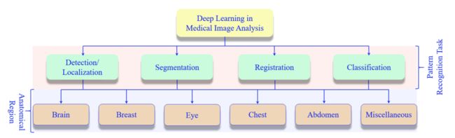
医学图像分析主要包含的模式识别任务是 检测/定位、分割、配准、分类。常见的医学影像包括 Brain、Breast、Eye、Chest、Abdomen等。
-
检测/定位

-
分割

-
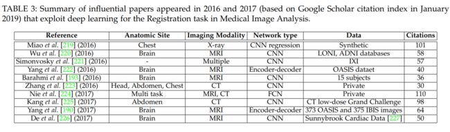
配准
-
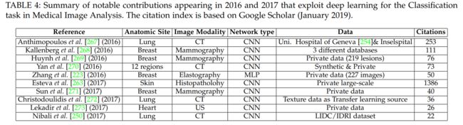
分类
Eye
GON
青光眼视神经病变 ScienceDirect paper
-
Li等[68]最近采用了深度转移学习方法,该方法可以很好地调整在ImageNet [69]数据集上预训练的VGG-16模型[31]。为了检测和分类眼中年龄相关性黄斑变性(AMD)和糖尿病性黄斑水肿(DME)疾病,他们使用207,130视网膜光学相干断层扫描(OCT)图像。该方法在视网膜图像中实现了98.6%的预测检测精度,100%。
-
Ambramoff等[70]使用基于CNN的技术检测眼底图像中的糖尿病视网膜病变(DR)。他们使用公共数据集[71]在他们的研究中评估了设备IDx-DRX2.1,并获得了0.98的AUC分数。
-
Schlegl等[72]采用深度学习方法检测视网膜图像中的视网膜内囊样液(IRC)和视网膜下液(SRF)。他们采用自动编码器-解码器形成CNN,并使用1,200个OCT视网膜图像进行实验,SRF的AUC为0.92,IRC的AUC为0.94。
-
Li等[75]基于Inception架构[43]训练了一个深度学习模型,用于识别视网膜图像中的青光眼视神经病变(GON)。他们的模型达到了0.986的AUC,用于区分健康眼睛和GON眼睛。
-
Christopher等[76]也使用VGG16,Inception v3和ResNet50模型进行转移学习来识别GON。他们使用预先训练的ImageNet模型。对于他们的实验,他们使用14,822个光学神经头(ONH)眼底图像的GON或健康的眼睛。据报道,在眼睛中鉴定中度至重度GON的最佳性能为AUC值0.97,灵敏度为90%,特异性为93%。 Khojasteh等[77]在DIARETDB1[78]和e-Ophtha[79]数据集上使用预先训练的ResNet-50检测视网膜图像中的出口。他们报告的准确度为98%,使用数据检测灵敏度为99%。
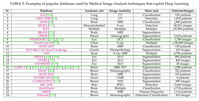
Datasets
Future
- 小数据问题:迁移学习、数据增广、GAN样本生成
- 图像结合病例资料的多模态学习
- 参考计算机视觉、机器学习领域的新工作
References
[43] C. Szegedy, W. Liu, Y. Jia, P. Sermanet, S. Reed, D. Anguelov,D. Erhan, V. Vanhoucke, and A. Rabinovich, “Going deeper with convolutions,” in Proceedings of the IEEE conference on computer vision and pattern recognition, 2015, pp. 1–9.
[75] Z. Li, Y. He, S. Keel, W. Meng, R. T. Chang, and M. He, “Efficacy of a deep learning system for detecting glaucomatous optic neuropathy based on color fundus photographs,” Ophthalmology,2018.
[76] M. Christopher, A. Belghith, C. Bowd, J. A. Proudfoot, M. H.Goldbaum, R. N. Weinreb, C. A. Girkin, J. M. Liebmann, and L. M. Zangwill, “Performance of deep learning architectures and transfer learning for detecting glaucomatous optic neuropathy in fundus photographs,” Scientific reports, vol. 8, no. 1, p.16685, 2018.
[77] P. Khojasteh, L. A. P. Junior, T. Carvalho, E. Rezende, B. Aliah- ´mad, J. P. Papa, and D. K. Kumar, “Exudate detection in fundus images using deeply-learnable features,” Computers in biology and medicine, vol. 104, pp. 62–69, 2019.
[78] R. Kalvi ¨ ainen and H. Uusitalo, “Diaretdb1 diabetic retinopathy ¨ database and evaluation protocol,” in Medical Image Understanding and Analysis, vol. 2007. Citeseer, 2007, p. 61.
[79] E. Decenciere, G. Cazuguel, X. Zhang, G. Thibault, J.-C. Klein, ` F. Meyer, B. Marcotegui, G. Quellec, M. Lamard, R. Danno et al.,“Teleophta: Machine learning and image processing methods for teleophthalmology,” Irbm, vol. 34, no. 2, pp. 196–203, 2013.
[128] J. Staal, M. D. Abramoff, M. Niemeijer, M. A. Viergever, and `B. Van Ginneken, “Ridge-based vessel segmentation in color images of the retina,” IEEE transactions on medical imaging, vol. 23, no. 4, pp. 501–509, 2004.
[129] M. M. Fraz, P. Remagnino, A. Hoppe, B. Uyyanonvara, A. R. Rudnicka, C. G. Owen, and S. A. Barman, “An ensemble classification-based approach applied to retinal blood vessel segmentation,” IEEE Transactions on Biomedical Engineering, vol. 59, no. 9, pp. 2538–2548, 2012.
[130] A. Hoover, V. Kouznetsova, and M. Goldbaum, “Locating blood vessels in retinal images by piecewise threshold probing of a matched filter response,” IEEE Transactions on Medical imaging, vol. 19, no. 3, pp. 203–210, 2000.
[168] P. Liskowski and K. Krawiec, “Segmenting retinal blood vessels with deep neural networks,” IEEE transactions on medical imaging, vol. 35, no. 11, pp.2369–2380, 2016.
[172] L. Fang, D. Cunefare, C. Wang, R. H. Guymer, S. Li, and S. Farsiu,“Automatic segmentation of nine retinal layer boundaries in oct images of non-exudative amd patients using deep learning and graph search,” Biomedical optics express, vol. 8, no. 5, pp. 2732–2744, 2017.
[276] G. Quellec, M. Lamard, P. M. Josselin, G. Cazuguel, B. Cochener, and C. Roux, “Optimal wavelet transform for the detection of microaneurysms in retina photographs.” IEEE Transactions on Medical Imaging, vol. 27, no. 9, pp. 1230–41, 2008.
[277] E. Decenciere, X. Zhang, G. Cazuguel, B. Lay, B. Cochener, `C. Trone, P. Gain, R. Ordonez, P. Massin, A. Erginay et al.,“Feedback on a publicly distributed image database: the messidor database,” Image Analysis & Stereology, vol. 33, no. 3, pp. 231–234,2014.
[279] M. Niemeijer, J. Staal, B. van Ginneken, M. Loog, and M. D.Abramoff, “Comparative study of retinal vessel segmentation methods on a new publicly available database,” in Medical Imaging 2004: Image Processing, vol. 5370. International Society for Optics and Photonics, 2004, pp. 648–657.