重要修复:Inobitec DICOM Viewer 2.11.1 Pro Crack
About the release of the Inobitec DICOM Viewer 2.11.1 Lite and Pro editions. Released on September 15, 2023.
History of changes:
Legend: [+] Addition, [*] Enhancement, [-] Elimination of a defect.
The main differences between the editions of Lite and Pro of version 2.11.0
Inobitec DICOM Viewer — software for visualization, archiving and exporting of medical images of DICOM format, obtained from medical equipment of various manufacturers.
- Perpetual licenses
- Multi platform support: Windows, macOS, Linux
- Does not impose high system requirements
- Provides advanced functionality for working in 3D
- Contains a detailed user manual in English
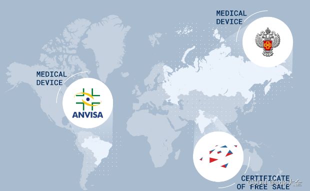
- Registered as a medical device in Roszdravnadzor
- Available in several editions, with discounts provided to distributors
Version 2.11.1
General changes for both editions:
- [*] Display of additional DICOM tags on ultrasound studies
- [-] Fixed several minor and critical bugs
DICOM Viewer Functionality
| Function | Lite | Pro | Web |
| View flat images with the following options: | ✔ | ✔ | ✔ |
| rotate, pan, zoom, mirror | ✔ | ✔ | ✔ |
| change window width and level | ✔ | ✔ | ✔ |
| change the window width and level based on the image parameters (Full dynamic W/L) | ✔ | ✔ | |
| customization of WL presets | ✔ | ✔ | |
| opening DICOM studies from zip archives | ✔ | ✔ | |
| opening several DICOM studies at the same time | ✔ | ✔ | ✔ |
| automatic filling tab windows with the series | ✔ | ✔ | |
| synchronization of series windows in case of window level/width alteration, movement, scaling, and scrolling | ✔ | ✔ | ✔ |
| calibrate image size | ✔ | ✔ | ✔ |
| show image scout lines on other series images | ✔ | ✔ | ✔ |
| flat image view with orthogonal projections | ✔ | ✔ | |
| measure length, angles | ✔ | ✔ | ✔ |
| play series images subsequently as a movie | ✔ | ✔ | ✔ |
| choose image resampling filter in the Flat Images View window | ✔ | ✔ | |
| build freeform contours with the Pencil tool | ✔ | ✔ | |
| add comments, labels and various graphic elements on images | ✔ | ✔ | ✔ |
| moving comments, measurements, measurement results and various graphic elements on images | ✔ | ✔ | ✔ |
| measure Cobb angles and intensity at a certain point or in a certain area | ✔ | ✔ | ✔ |
| export images to a graphic file | ✔ | ✔ | |
| export images to a new series of DICOM images | ✔ | ✔ | ✔ |
| DSA (Digital Subtraction Angiography) | ✔ | ✔ | |
| measurement of speed and time interval for dopplergram | ✔ | ✔ | |
| distance measurement on ultrasound studies | ✔ | ✔ | |
| support for color lookup tables (CLUTs) embedded in DICOM data | ✔ | ✔ | |
| applying different CLUTs to images | ✔ | ✔ | ✔ |
| edit CLUTs | ✔ | ✔ | |
| browse DICOM image tags | ✔ | ✔ | ✔ |
| cerebral Perfusion | ✔ | ||
| blood flow parameters evaluation on the basis of phase-contrast images | ✔ | ||
| calcium scoring | ✔ | ||
| measurement of apparent diffusion coefficient (ADC) | ✔ | ||
| View three-dimensional tissue reconstruction with the following options: | ✔ | ✔ | ✔ |
| rotate, pan and zoom the model | ✔ | ✔ | ✔ |
| view scaled-down multiplanar reconstruction of the series simultaneously (with automatic synchronization) | ✔ | ✔ | |
| cut the outside and inside parts of the model | ✔ | ✔ | ✔ |
| MIP (Maximum Intensity Projection) | ✔ | ✔ | |
| measure length, angles | ✔ | ✔ | |
| export the model to a graphic file | ✔ | ✔ | |
| export the model to a new series of DICOM images | ✔ | ✔ | ✔ |
| export of several images obtained by rotating the model to a graphic file | ✔ | ✔ | |
| export of several images obtained by rotating the model to a new series of DICOM images | ✔ | ✔ | |
| switching projection | ✔ | ✔ | |
| add markers and line markers | ✔ | ✔ | |
| delete bone tissue in the manual mode (interactively and automatically) | ✔ | ✔ | |
| delete bone tissue for merged series | ✔ | ||
| build the surface by the model | ✔ | ||
| surface construction for the main volume | ✔ | ||
| surface building for each phase of the main volume in multiphase series | ✔ | ||
| individual change of parameters for each constructed surface | ✔ | ||
| measure the shortest path on the surface of a visible volume with the Surface Ruler tool | ✔ | ||
| import of multiple surfaces | ✔ | ||
| segmentation | ✔ | ||
| View multiplanar reconstruction with the following options: | ✔ | ✔ | ✔ |
| view axial, frontal and sagittal sections of tissue | ✔ | ✔ | ✔ |
| rotate cutting planes in space | ✔ | ✔ | ✔ |
| 3D view | ✔ | ✔ | ✔ |
| measure length, angles | ✔ | ✔ | ✔ |
| add markers and line markers | ✔ | ✔ | |
| build freeform contours with the Pencil tool | ✔ | ✔ | |
| setting modes MIP, minIP, AIP | ✔ | ✔ | ✔ |
| set slice thickness | ✔ | ✔ | ✔ |
| MPR view sync on several monitors | ✔ | ✔ | |
| display of MPR planes on 3D view | ✔ | ✔ | |
| building a curvilinear reconstruction | ✔ | ✔ | |
| build a section of a spatial model by a random surface | ✔ | ||
| cross-section view for curvelinear reconstruction | ✔ | ||
| export sections of any of the planes with a selectable increment to a series | ✔ | ||
| segmentation | ✔ | ||
| surface construction for the main volume | ✔ | ||
| surface building for each phase of the main volume in multiphase series | ✔ | ||
| individual change of parameters for each constructed surface | ✔ | ||
| import of multiple surfaces | ✔ | ||
| contour based segmentation | ✔ | ||
| showing the mask outline | ✔ | ||
| histogram display for ROI | ✔ | ||
| mixed RGB colors Fusion | ✔ | ||
| Diffusion Tensor Imaging (DTI) support | ✔ | ||
| Virtual endoscopy (viewing the inner surfaces of cavities in tissues) with the following options: | ✔ | ||
| automatically and manually navigate the inside a cavity | ✔ | ||
| build cavity surfaces | ✔ | ||
| view scaled-down multiplanar reconstruction of the series simultaneously (with automatic synchronization) | ✔ | ||
| Merge series with the following options: | ✔ | ||
| remove bone tissue for DualScan and DualEnergy studies | ✔ | ||
| build three-dimensional models based on multiple series of images of the same tissue in different modes | ✔ | ||
| quickly switch to a full-scale view of three-dimensional reconstruction, multiplanar reconstruction and virtual endoscopy of the model built by merging series | ✔ | ||
| stitch images | ✔ | ||
| View electrocardiogram with the following options: | ✔ | ✔ | ✔ |
| measure time intervals and values on graphs | ✔ | ✔ | ✔ |
| using filters | ✔ | ✔ | |
| export images to a new series of DICOM images | ✔ | ✔ | |
| Print images on paper or film using a DICOM printer with the following options: | ✔ | ✔ | |
| adding reference images | ✔ | ✔ | |
| exporting pages to the PACS server for printing | ✔ | ✔ | |
| adding logo | ✔ | ✔ | |
| Open studies by dragging and dropping on the program icon and program window | ✔ | ✔ | |
| Open studies in the background | ✔ | ✔ | |
| Transformation indicators in the series window with an opportunity to undo the transformations | ✔ | ||
| Open/save file using Native OS dialogs | ✔ | ✔ | |
| Voxel and polygon rendering support on Metal for macOS starting with Version 10.15 | ✔ | ✔ | |
| Set up the interface and the DICOM Viewer functionality with the capability to export and import the settings configuration | ✔ | ✔ | |
| Tool control with the left, the right and the middle mouse buttons | ✔ | ✔ | ✔ |
| High resolution monitor support | ✔ | ✔ | ✔ |
| The support of secure connection with TLS | ✔ | ✔ | |
| Viewing DICOM tags | ✔ | ✔ | ✔ |
| PDF data support and presentation in DICOM files | ✔ | ||
| Structured reports support and presentation | ✔ | ||
| The opportunity to view several types of data in one window simultaneously: flat images, volume reconstructions, MPR, ECG, video, PDF documents and structured reports | ✔ | ||
| Minimize application window to system tray | ✔ | ✔ | |
| Integrated help system | ✔ | ✔ | ✔ |
| Create series display templates for various modalities | ✔ | ✔ | |
| Write data to CD, DVD and flash cards | ✔ | ✔ | |
| Integration with PACS servers | ✔ | ✔ | ✔ |
| Store data in a local storage | ✔ | ✔ | ✔ |
| Work as a PACS server, support C-FIND and C-MOVE | ✔ | ✔ | |
| Saving studies to a folder | ✔ | ✔ | ✔ |
| Saving selected series to a folder | ✔ | ✔ | |
| Integration with third-party systems | ✔ | ✔ | ✔ |
| Edit patient name and study description on the Local Storage | ✔ | ✔ | |
| Anonymize studies and series with the ability to change DICOM tags | ✔ | ✔ | |
| PLY, OBJ, STL, GLB formats surface export | ✔ | ||
| PLY, OBJ, STL formats surface import | ✔ | ||
| Saving the model orientation and projection in spj-projects | ✔ | ||
| Embedded WebViewer | ✔ | ||
| Register images | ✔ | ||
| Record video from viewing windows | ✔ | ||
| Play video contained in DICOM files | ✔ |


