Part 1 Artificial Neural Networks(ANN)
Topic 1 Historical/Biological Introduction
1. Biological Excitability
(a. Virtually all living cells maintain an electrical potential difference between their
interiors and the environment (exteriors) . 内部和外部环境存在电压差
(b. The membrane potential(膜电位) is one of the factors determining the energy barriers
encountered by charged substances (ions) entering or leaving the cell
(c. Within the cell membrane there are ion channels(离子通道) – proteins with the central(带有中心孔的蛋白质) pore through which ions can cross the membrane
(d. A schematic diagram of a section of the lipid bilayer(脂质双分子层) that forms the cell membrane with two ion channels embedded in it. The membrane is 3 to 4 nm thick, and the ion channels are about 10 nm long.

1. Under resting conditions ( normal, inactive, not sending a signal ), the potential inside neuron membrane is from -30 mV to -90 mV relative to that of the surrounding bath, conventionally defined to be 0 mV, and the cell is said to be polarized .
在静息条件下(正常、不活跃、不发送信号),神经元膜内的电位相对于周围浴液的电位从-30 mV到-90 mV,通常定义为0 mV,细胞被极化。
2. The electrical signal of a living cell is change of the membrane potential .
活细胞的电信号是膜电位的变化。
3. The membrane potential may change in response to electrical perturbation from other neurons.
4. If the perturbation is sufficiently large, above a threshold in intensity and duration , the response is a large amplitude electrical wave, propagating from the stimulated points to the rest of the tissue.
如果扰动足够大,强度和持续时间超过阈值,则响应是一个大振幅大的电波,从受刺激点传播到组织的其他部分。
Wave Propagation
1. The wave travels with a nearly uniform velocity .匀速
2. The excitation and transmission are all-or-none and do not allow varying
degree of strength.
激发之后是一段有一定持续时间的不可兴奋期,称为绝对不应期
3. Excitation is followed by an unexcitable period of definite duration , called the
absolute refractory period; which is followed in turn by a relative refractory
period when the cell has subnormal excitability.
1. Important morphological specializations of neurons are the dendrites that receive inputs from other neurons, soma (the cell body), and the axon that carries the neuronal output to other cells.
2. The elaborate branching structure of the dendritic tree allows a neuron to receive inputs from many other neurons through synaptic connections.
神经元重要的形态学特化是接收来自其他神经元输入的树突、体细胞(细胞体),以及将神经元输出传递到其他细胞的轴突。树突状树的复杂的分支结构允许一个神经元通过突触连接接收来自许多其他神经元的输入。
The dendrites and soma act as input surface for signals from other neurons and/or receptors.
The axon carries signals from the neuron to other neurons and/or effectors (e.g., muscle fibers or glands)
树突和体细胞作为来自其他神经元和/或受体的信号的输入表面。轴突将信号从神经元传递到其他神经元和/或效应器(如肌纤维或腺体)
• (A): cortical pyramidal cell. These are the primary excitatory neurons of the cerebral cortex. Pyramidal cell axons branch locally, sending signals to synapse with nearby neurons, and also more distally to other parts of the brain and nervous system.
• (B): Purkinje cell. Purkinje cell axons transmit the output of the cerebral cortex.
• (C): stellate cell. Stellate cells are one of a large class of interneurons that provide inhibitory input to the neurons of the cerebral cortex.
(A):皮质锥体细胞。这些是大脑皮层的主要兴奋性神经元。锥体细胞轴突在局部分支,向附近神经元的突触发送信号,也更远端到大脑和神经系统的其他部分。•(B):浦肯野细胞。浦肯野细胞轴突传递大脑皮层的输出。•(C):星状细胞。星状细胞是为大脑皮层的神经元提供抑制性输入的大量中间神经元之一。
The cortical pyramidal neuron A and the cortical interneuron C each receive thousands of synaptic inputs, and for the Purkinje cell B, the number is over 100,000.
• Axons from single neurons can traverse large fractions of the brain or, in some cases, of the entire body.
• Axons can also connect with multiple targets.
皮层锥体神经元A和皮层间神经元C分别接收数千个突触输入,而对于浦肯野细胞B,其数量超过10万个。来自单个神经元的 •轴突可以穿过大脑的大部分,在某些情况下,还可以穿过整个身体。轴突也可以与多个目标相连接
The tips of the axon branches are called nerve terminals or boutons .
• The location of interaction between a terminal and the cell upon is called a synapse .
• A synapse shown in the left figure illustrates the presynaptic bouton at the end of the axon and a spine on the dendrite.
轴突分支的尖端被称为神经末梢或钮扣。•终端和细胞之间相互作用的位置称为突触。•左图所示的一个突触显示了轴突末端的突触前钮扣和树突上的一个脊柱。
The terminology of presynaptic and postsynaptic defines the direction of signal flow.
• Santiago Ramon y Cajal, ~1901, supposed that the specific networking of the nervous cells determines direction of transmission of information . This discovery made clear that the coupling of the neurons constitutes a hierarchical system .
突触前和突触后的术语定义了信号流的方向。•圣地亚哥Ramon y Cajal,~1901,认为神经细胞的特定网络决定了信息传递的方向。这一发现清楚地表明,神经元的耦合构成了一个层次系统。
The chemical transmission of information at the synapses was mostly studied from 1920 to 1940.
• The two neurons are not directly connected but communicate via the cleft .
突触上信息的化学传递主要是在1920年至1940年期间进行研究的。•这两个神经元不是直接连接的,而是通过裂缝进行交流。
The axon terminal or bouton is filled with synaptic vesicles containing neurotransmitter .
• The neurotransmitter is released when a spike arrives from the presynaptic neuron
轴突末端或钮扣上充满了含有神经递质的突触囊泡。•当突触前神经元到达时,神经递质被释放
Transmitter crosses the synaptic cleft and binds to receptors on the dendritic spine.
• Excitatory synapses on cortical pyramidal cells form on dendritic spines as shown here. Synapses can form on the dendrites, or axon.
传递器穿过突触间隙并与树突棘上的受体结合。•皮质锥体细胞上的兴奋性突触形成于树突状棘上,如图所示。突触可以在树突或轴突上形成。
Although impulses spread uniformly along axons, there is no physiological continuity from neuron to neuron .
• When an impulse (perturbation) reaches a synapse, it does not necessarily stimulate the following neuron .
虽然脉冲沿轴突均匀扩散,但神经元之间没有生理连续性。•当一个脉冲(扰动)到达一个突触时,它并不一定会刺激以下的神经元。
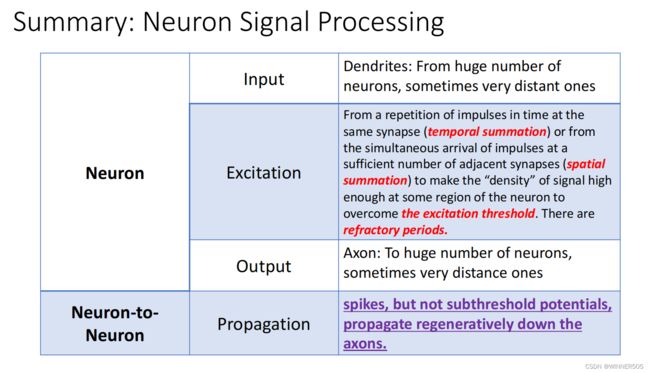
Trans-synaptic stimulation of a neuron requires usually
• either a repetition of impulses in time at the same synapse ( temporal summation ).
• or the simultaneous arrival of impulses at a sufficient number of adjacent synapses ( spatial
summation )
to make the “density” of excitation high enough at some region of the neuron.
神经元的跨突触刺激通常需要在同一突触上重复时间脉冲(时间总和)。•或脉冲同时到达足够数量的相邻突触(空间总和),使神经元的某些区域的兴奋“密度”足够高。
The arrival of impulses at synapses may have opposite to excitation effect, i.e., it may render the element less excitable to other stimuli. This decrease of excitability is called inhibition .
脉冲在突触上的到来可能与兴奋效应相反,也就是说,它可能使该元素对其他刺激不那么兴奋。这种兴奋性的降低被称为抑制
1. The excitatory or inhibitory effect of the transmitter generally causes a potential change in the postsynaptic membrane.
2. The cooperative effect of many potential changes may yield a synthesized potential change in the soma that exceeds the threshold – and if this occurs at a time
3. when the neuron has passed the refractory period of its previous firing, then a new impulse is fired down the axon.
递质的兴奋性或抑制性作用通常会引起突触后膜的潜在变化。•许多潜在的变化的合作效应可能产生一个合成潜在的躯体变化超过阈值,如果这发生在神经元通过的耐火的不应期,然后一个新的脉冲发射轴突。
The top trace represents a recording from an intracellular electrode connected to the soma of
the neuron. The height of the action potentials has been clipped to show the subthreshold membrane potential more clearly.
• The middle trace is a simulated extracellular recording . Action potentials appear as roughly
equal positive and negative potential fluctuations with an amplitude of ~0.1mV, which is ~1000 times smaller than the approximately 0.1V amplitude of an intracellularly recorded action potential.
• The bottom trace represents a recording from an intracellular electrode connected to the axon some distance away from the soma. The full height of the action potentials is indicated in this trace.
顶部的痕迹代表了一个连接到神经元体细胞的细胞内电极的记录。裁剪了动作电位的高度,以更清楚地显示阈下膜电位。•中间的痕迹是一个模拟的细胞外记录。动作电位表现为大致相等的正负电位波动,振幅为~0.1mV,比细胞内记录的动作电位的约0.1V振幅小1000倍。•底部的痕迹代表了一个在离躯体一定距离处连接到轴突的细胞内电极的记录。动作电位的全高度在这个轨迹中表示。
The top recording from soma shows rapid spikes riding on top of a more slowly varying subthreshold potential.
• The bottom trace shows intracellular recording from axon some distance out of soma. The
subthreshold membrane potential waveform, apparent in the soma recording, is completely absent on the axon due to attenuation, while the action potential sequence in the two recordings is the same.
• The difference in records from soma and from the axon illustrates the important point that
spikes, but not subthreshold potentials, propagate regeneratively down the axons.
来自躯体的顶部记录显示,快速的峰值骑在一个更缓慢变化的阈下电位之上。•底部的痕迹显示了从轴突离体细胞有一定距离的细胞内记录。阈下在体细胞记录中明显的膜电位波形由于衰减在轴突上完全缺失,而两个记录中的动作电位序列相同。•来自体细胞和轴突的记录的差异说明了重要的一点,即峰值,而不是阈下电位,沿着轴突再生传播。


